Osteochondritis dissecans
Osteochondrosis dissecans, cartilage damage, König's disease
Osteochondritis dissecans is a disorder of the cartilage and underlying bone of a joint. As a result, a piece of bone with cartilage can become detached from the joint surface.

A loose piece of tissue in a joint is also called a corpus liberum. It can cause pain and mobility problems in the joint. The knee is by far the most frequently affected area, but it can also occur in other joints.
Description of the condition
In osteochondritis dissecans, cartilage is damaged in combination with the bone directly beneath it. The bone is the first to deteriorate and, at some point, dies.
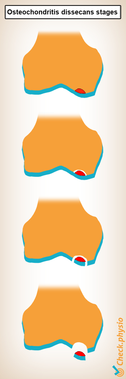
At a later stage, a piece of bone, together with a piece of cartilage, can break off and end up in the joint. In this respect, it can start to 'float around' and cause joint locking as a result of which a joint locks up, as it were, when the patient tries to move. The cartilage damage itself can also cause (painful) symptoms during movement.
Osteochondritis dissecans is more common in boys than in girls. Especially when exercising intensively. This disorder occurs most frequently between the ages of 10 and 20. However, it can also affect people later in life. That is why there is a juvenile and an adult form.
The juvenile form means that the bones are not yet fully grown. This form is easier to treat than in adults because the recovery capacity of cartilage is better at a younger age.
Cause and origin
The cause of osteochondritis dissecans is not yet known. However, there are a number of theories that offer a possible explanation. For example, genetic predisposition, poor circulation, gender, overexertion or (repeated) trauma (accident/fall) play a role. It is probably a combination of the above factors that ultimately leads to osteochondritis dissecans.
People with osteochondritis dissecans often have damage in several joints. This damage occurs particularly in the joints that are subjected to greater strain during sport or work.
Stages
Osteochondritis dissecans is divided into stages that increase in severity. The first stage starts with the death of the bone below the cartilage surface, without affecting the cartilage. At that time, there are usually no symptoms yet. As the condition worsens, bone and cartilage become detached.
In the final stage, there is complete detachment with displacement of one or more pieces of bone and cartilage. This is referred to as a corpus liberum and severe joint damage.
Signs & symptoms
People with osteochondritis dissecans may experience the following symptoms to varying degrees:
- Pain when straining the affected joint.
- Swelling of the joint.
- Joint locking (the joint locks up when making a movement).
- Feeling unsteady.
- Limited movement of the affected joint.
Diagnosis
A doctor or physiotherapist will inquire about the symptoms and how they arose. Afterwards, a physical examination will be performed to check for any swelling, joint locking and mobility of the knee.
If osteochondritis dissecans is suspected, the first step is to take an X-ray. This will show the condition, particularly in its advanced stages. If the X-ray does not show anything obvious, an MRI can be taken. This will also show any abnormalities in the joint at an early stage. In addition, an MRI makes it easier to differentiate between osteochondritis dissecans and other disorders with the same complaints.
Treatment
Treatment depends on the patient's age and the stage of the disease. When a person develops osteochondritis dissecans at a young age, and it is caught relatively early, a wait-and-see approach is sufficient. In this case, the weight-bearing of the joint depends on the symptoms so that the joint can heal by itself. If a piece of bone and cartilage has already come loose, surgery will follow.
Surgery
When an older person has complaints related to osteochondritis dissecans, surgery is almost always required. This is because spontaneous recovery is rare. This surgical procedure also depends on the stage the condition is in. For example, a somewhat larger fragment can be placed back in its place with a screw. Another way is to take pieces of bone and cartilage from another part of the body and place them in the damaged area. This creates a kind of mosaic pattern.
Rehabilitation
After the operation, a rehabilitation period with a physiotherapist follows. This takes about three to six months. The weight-bearing on the joint is gradually increased while focussing on strength, stability, mobility and stamina.
The result of the treatment depends on several factors. Age, weight, size of the damage and the stage of the disease play a major role.
Exercises
You can check your symptoms using the online physiotherapy check or make an appointment with a physiotherapy practice in your area.
References
Jafari D., Shariatzadeh H., Mazhar F.N., Okhovatpour M.A. & Razavipour, M. (2017). Osteochondritis dissecans of the humeral Head. A case report and review of the literature. Arch Bone Jt Surg. 2017; 5(1): 66-69.
Magee, D.J., Zachazewski, J.E., Quillen, W.S., Manske, R.C. (2016). Pathology and intervention in musculoskeletal rehabilitation. Elsevier, 2nd edition.
Nugteren, K. van & Winkel, D. (2008). Onderzoek en behandeling van de knie. Houten: Bohn Stafleu van Loghum.
Verhaar, J.A.N. & Linden, A.J. van der (2005). Orthopedie. Houten: Bohn Stafleu van Loghum.


